Why Does Posterior Acoustic Enhancement Appear Bright? Ultrasound Artifacts | Fluid Filled Spaces

Why Does Posterior Acoustic Enhancement Appear Bright? Ultrasound Artifacts | Fluid Filled Spaces
Sound Wave Transmission: When an ultrasound beam travels through the body, it encounters various tissues and structures, each with different acoustic properties.
Some tissues attenuate (absorb and scatter) the ultrasound waves more than others. For instance, fluids (like cysts or bloodfilled structures) have low attenuation compared to solid tissues (like muscles or organs).
Low Attenuation Structures: Structures such as cysts or fluidfilled spaces allow ultrasound waves to pass through with minimal attenuation.
As the sound waves pass through these low attenuation areas, they lose less energy compared to the surrounding tissues.
After passing through the low attenuation area, the sound waves reach the tissues behind it (posterior to the fluidfilled structure) with more intensity than if they had traveled through more attenuating tissues.
The ultrasound machine then receives stronger echoes from the tissues behind the low attenuation structure.
Fluid in a cyst, has low attenuation compared to solid liver tissue.
As the ultrasound wave passes through the cyst, it loses very little energy because fluid does not significantly absorb or scatter the sound waves.
After passing through the cyst, the ultrasound wave reaches the solid liver tissue that lies posterior (behind) the cyst.
The ultrasound waves reflect off the solid liver tissue behind the cyst with relatively more energy than they would if they had traveled through solid liver tissue all along. These stronger echoes are picked up by the transducer.
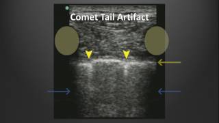
The ultrasound machine processes these stronger returning echoes and represents them as an area of increased brightness or echogenicity on the image. This increased brightness or echogenicity behind the cyst is known as posterior acoustic enhancement.
The cyst itself appears as an anechoic (dark) area because fluid does not produce many echoes. Directly behind this anechoic area, there is a region of increased brightness, indicating posterior acoustic enhancement.
The vitreous humor is a gellike fluid that fills the posterior chamber of the eye. Similar to other fluidfilled structures, the vitreous humor has low attenuation.
A simple breast cyst is a fluidfilled sac within the breast tissue. Fluids have low attenuation properties, meaning they do not significantly absorb or scatter ultrasound waves.
An abscess is a localized collection of pus, which consists of fluid, cellular debris (such as dead white blood cells, bacteria, and tissue cells), and other inflammatory materials. The mixture of fluid and solid particles within the abscess creates a heterogeneous internal structure. As the ultrasound waves pass through the abscess, they encounter both fluid and cellular debris.
The fluid component of the abscess attenuates the ultrasound waves very little, allowing most of the wave's energy to continue through the fluid. The cellular debris and other solid components within the abscess can reflect and scatter some of the ultrasound waves. Despite some reflection from cellular debris, the fluid component still allows a significant portion of the ultrasound waves to pass through with minimal attenuation.